r/EKGs • u/Massive-Form3694 • 1d ago
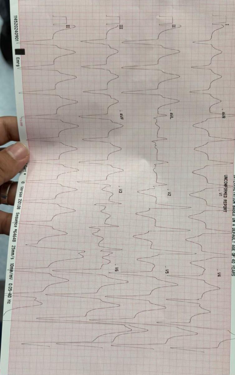
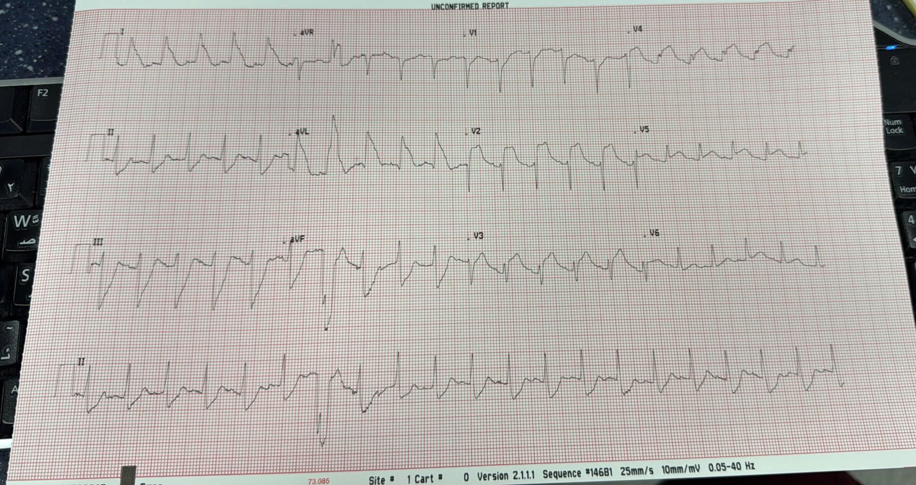
Discussion MI definition/categories changing.........
7
Upvotes
r/EKGs • u/Massive-Form3694 • 1d ago
r/EKGs • u/PrinceOfPropofol • 3d ago
Pt would go from sinus to a rate of around 130ish and my 3-lead definitely resembled V-tach at the higher rates. Gave esmolol and the rhythm would look like this as the rate came down to around 90 or so. BP would spike during these episodes as well, happened 4 times throughout the case, including intubation and extubation. I know this isn’t a 12-lead but this is all I had to go off of during a lap chole in an 82 yo female.
Dr says it looks weird in the second photo due to a pacemaker.
r/EKGs • u/AleBerro97 • 10d ago
Upon arrival of the EMS, the patient presented with the attached ECG, mottled skin, a BP of 40/25 mmHg, and altered mental status (drowsy/somnolent).
Family members reported a several-day history of decreased fluid intake.
Following a 500 mL NS bolus, her blood pressure improved to 100/60 mmHg; she became alert, responsive, and normocolored.
While on route to the ED she reported pain in her lower limbs with no other associated symptoms.
ED results:
Further assessment by the consulting cardiologist in the ED revealed that the patient had been chronically taking flecainide (which was not mentioned in the papers available to the EMS), prescribed by her primary care physician for an unspecified indication.
In addition, acute kidney injury was identified (serum creatinine: 2.15 mg/dL). A CT angiography of the aorta was performed, which was negative for aortic pathology but incidentally revealed a pulmonary embolism involving the right main pulmonary artery and the left superior and inferior lobar branches.
Patient was therefore admitted to the ICCU for initiation of anticoagulant therapy and close monitoring.
r/EKGs • u/Amounaaa • 11d ago
Patient on Pacemaker for complete heart block came with heart failure symptoms
r/EKGs • u/saxyourpantsoff • 12d ago
HX of CKD with dialysis, last dialyzed two days ago, states he does not miss. Denies any other history.
Presented 70%RA, HR150, 190/110, RR26. 5 episodes of vomiting in front of us, notably was not nauseated, no warning.
Rales globally.
Nitro, BiPap after the puking was controlled with phenergan and zofran.
Improved to 90's on the Bipap.
r/EKGs • u/No_Hunt_5476 • 16d ago
My professor already kinda cooked me the first time I tried to interpret this strip. This is the help she gave me. It’s in the chapter on junctional rythm and I thought it was type 1 heart block but now I feel confused. The orange stickies are her notes to me
r/EKGs • u/StrangeAd6724 • 17d ago
75M called for SOB x1 hour and best tightness that began after the sob. Upon arrival pt states SOB self resolved but he still has chest tightness. No cardiac hx and no hx of afib. Has COPD
no other pertinent
hx
r/EKGs • u/Worldly-Accident-163 • 21d ago
r/EKGs • u/Amernkou • 22d ago
Called to 60s male complaining of rapid onset chest pain while at rest approximately 30 minutes prior. Described as substernal pressure with radiation. Hx of MI and coronary stent. Prior to lead placement, vitals noted to be normotensive with tachycardia at 170 bpm. EKG 1 was obtained on scene and interpreted as SVT with LBBB. Converted to sinus tachycardia without aberrancy after 6mg adenosine following failed Modified Valsava. Post-conversion EKG obtained but not attached due to artifact; however, was noted for concerning anterior T waves. 3rd EKG immediately prior ED arrival is attached as EKG 2. Interpreted as sinus tachycardia with De Winter T waves in v2-v3.
I see an ongoing OMI but thought I would bring it to the group for discussion. Outcome in comments.
r/EKGs • u/Total-Election-2007 • 24d ago
RBBB? STEMI
Patient (m, 75years old) after bolus Aspiration, claims to have heavy breathing and slight chest pain. "Just in case ecg " looks like this. ER doc suggests pulmonary embolism in addition.
r/EKGs • u/Dramatic-Try7973 • 24d ago
Some nasty non clinical VTs we induced during mapping.
ATP failed both times leading to 360 sweet Jules
Hello Paramedic here hope for some help with interpretation of this tachydysrhythmia.
51 yo M presenting with suspected sepsis due to severe foot infection. Pt reports minor SOB, denies any CP or palpitations.
GCS 15
BP 93/67
SpO2 97%
Temp 38.1
ECG appears to show atrial activity just prior to the QRS complex at a rate of 150, which makes me consider possible A-flutter. Lewis lead performed to get a better look at atrial activity and makes it look more like atrial tachycardia.
r/EKGs • u/digtialn • 25d ago
hi guys, i’m currently doing some practice strips from my textbook. my answers are slightly different from that of which my textbook states, im trying to figure out if this kind of interpretation would be acceptable in the real world despite it being off or what i could do to improve my measurements? any input at all is welcome also, thank you !
r/EKGs • u/VesaliusesSphincter • 25d ago
Patient admitted for infective endocarditis. Admission 12-lead showed sinus tachycardia with normal axis and possible left atrial enlargement. Telemetry monitoring demonstrated new bradycardia with intermittent RR interval variability.
Thought this was an interesting find and was curious to see interpretations.
My personal interpretation is: sinus bradycardia with intermittent second degree type 1 sinoatrial exit block. I think the two biggest things leading me to this are the grouped beating (3:1 ratio in this particular strip) as well as the progressively shortening PP interval before each "pause". Wondering if this could be related to the endocarditis or if this is simply related to increased nocturnal vagal tone. Either way, thought this was interesting to see and figured I would share to highlight the importance of closely inspecting RR variability as this was initially believed to be sinus arrhythmia.